Miles de libros con descuentos a un sólo clic - 50% de dto  Ver más

Enviar a
Montevideo, Montevideo
0
  • argentina
  • chile
  • colombia
  • españa
  • méxico
  • perú
  • estados unidos
  • internacional

Selecciona tu país

América

Europa

Resto del mundo

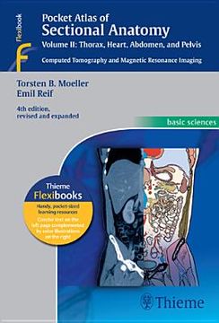
portada pocket atlas of sectional anatomy, volume ii: thorax, heart, abdomen and pelvis: computed tomography and magnetic resonance imaging (en Inglés)
Formato
Libro Físico
Año
2013
Idioma
Inglés
N° páginas
346
Encuadernación
Tapa Blanda
Dimensiones
18.8 x 12.7 x 2.0 cm
Peso
0.48 kg.
ISBN
3131256044
ISBN13
9783131256041
N° edición
0004

pocket atlas of sectional anatomy, volume ii: thorax, heart, abdomen and pelvis: computed tomography and magnetic resonance imaging (en Inglés)

Torsten Bert Moeller (Autor) · Torsten Bert Möller (Autor) · Emil Reif (Autor) · Thieme Medical Publishers · Tapa Blanda

pocket atlas of sectional anatomy, volume ii: thorax, heart, abdomen and pelvis: computed tomography and magnetic resonance imaging (en Inglés) - Moeller, Torsten Bert ; Möller, Torsten Bert ; Reif, Emil

Libro Nuevo Importado
Envío: 17 a 24 días háb.
$ 5.057$ 2.529
-50%
Costos de importación incluídos en el precio ✅
Libro Nuevo

Quedan 2 unidades

$ 2.529
Llega entre el 18 Ago y el 28 Ago a Montevideo, Montevideo. Seleccionar ubicación

Reseña del libro "pocket atlas of sectional anatomy, volume ii: thorax, heart, abdomen and pelvis: computed tomography and magnetic resonance imaging (en Inglés)"

This comprehensive, easy-to-consult pocket atlas is renowned for its superb illustrations and ability to depict sectional anatomy in every plane. Together with its two companion volumes, it provides a highly specialized navigational tool for all clinicians who need to master radiologic anatomy and accurately interpret CT and MR images.Special features of Pocket Atlas of Sectional Anatomy: Didactic organization in two-page units, with high-quality radiographs on one side and brilliant, full-color diagrams on the other Hundreds of high-resolution CT and MR images made with the latest generation of scanners (e.g., 3T MRI, 64-slice CT) Color-coded schematic drawings that indicate the level of each section Consistent color coding, making it easy to identify similar structures across several slicesUpdates for the 4th edition of Volume II: CT imaging of the chest and abdomen in all 3 planes: axial, sagittal, and coronal New back-cover foldout featuring pulmonary and hepatic segments and lymph node stations Follows standard international classifications of the American Heart Association for cardiac vessels and the AJCC/UICC for mediastinal lymph nodes Compact, easy-to-use, highly visual, and designed for quick recall, this book is ideal for use in both the clinical and study settings.

Opiniones del libro

Preguntas frecuentes sobre el libro

Todos los libros de nuestro catálogo son Originales.
El libro está escrito en Inglés.
La encuadernación de esta edición es Tapa Blanda.

Preguntas y respuestas sobre el libro

¿Tienes una pregunta sobre el libro? Inicia sesión para poder agregar tu propia pregunta.

Opiniones sobre Buscalibre

Ver más opiniones de clientes